You are here > Home / Radiology
What is Radiology ?
Radiology, also known as diagnostic imaging, is a series of tests that takes images of several body parts. It is a medical branch that deals with the usage of controlled beams of intense energy in the diagnosis as well as the treatment of various diseases. Many such tests are unique as they allow doctors to check the inner conditions of the body. Radiology is vital to the diagnosis of various diseases, specifically cancers. Early diagnosis through radiology can save lives. Radiologists are physicians who specialize in interpreting such imaging examinations’ results. It includes several tests like digital x-ray, ultrasonography, echocardiography, CT, MRI, and many more.
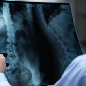
Digital X-Ray (DR Imaging System)
Digital x-ray, also known as digital radiography, is a type of imaging that uses sensors of digital x-rays instead of a traditional photo film. As it uses a digital device to capture images, it allows the data to get transferred to a laptop, desktop, or any other digital medium without utilizing an intermediate medium. Digital x-ray produces small radiation and provides a high-resolution image in a digital x-ray centre that can be enlarged as per requirement. Digital x-ray system also uses virtual storage, which is easy to access.

Ultra Sonography
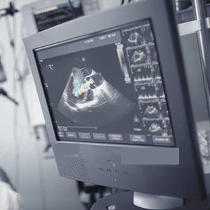
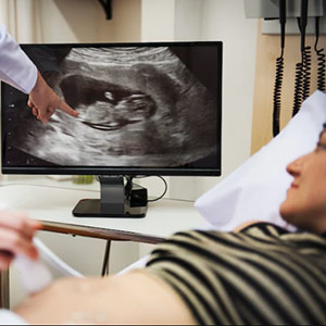
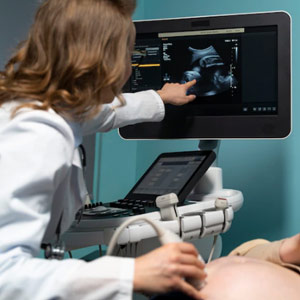
Ultrasonography test is an imaging technology that transfers high-frequency sound waves to look at tissues and organs inside the body. With the help of ultra-sonography, an echo signal is produced, which is captured by a desktop or a laptop to create a real-time representation of the anatomy and physiology of the body. This imaging approach is the least intrusive of all the options available to make it more acceptable for usage, especially during a delicate situation such as pregnancy. The best ultrasonography center in Kolkata uses this method to check the blood flow in the neck, heart function, and extremities, along with a few medical disorders.
Echo Cardiography
An eco-cardiography is a diagnostic imaging test that uses sound waves to produce images of your heart. It is one of the common tests through which doctors can see and check your heart, and it’s functions. With the help of eco-cardiography, a doctor can detect and identify heart diseases. Heart diseases like problems with the chambers or valves of the heart and congenital heart defects can also be detected through eco-cardiography. Echo-cardiography is a vital tool used to assess unusual wall motion among patients with heart issues. It can also detect cardiomyopathies like dilated cardiomyopathies, hypertrophic cardiomyopathies, and much more.

Doppler Study
A doppler study, also known as a doppler ultrasound, is a non-invasive examination that can predict blood flow through the blood vessels by bouncing high-frequency sound waves. With the help of a doppler study, conditions such as blood clots, poor functioning of valves, blockage in the artery, reduced blood circulation, bulging arteries, and many other issues can be detected. This examination is also done as an alternative to invasive procedures like angiography. Doctors use the doppler study to check for injuries to your arteries and monitor specific treatments for your arteries and veins.
CT
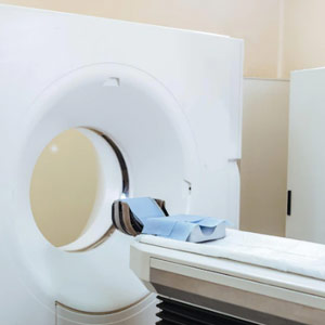
CT scan, or a computerized tomography scan, is a technique used for medical imaging. It is used for obtaining detailed internal pictures of the body. CT scan combines a series of X-ray pictures captured from various angles around the body. It uses computer processing to make cross-sectional pictures or slices of blood vessels, bones, and soft tissues in the body. A CT scan can also be used for various aspects like visualizing almost every body part and diagnosing diseases or injuries (mainly internal injuries). It takes less time to execute the diagnosis process and prepares the result quickly.
MRI
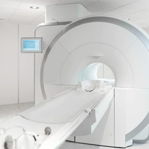
MRI, or magnetic resonance imaging, is a diagnostic imaging technique used to capture pictures of the physiological process and anatomy of the body. It works with the help of computer-generated radio waves to make detailed pictures of the organs and tissues in the body. Most machines for MRI are usually large and tube-shaped magnets. With the help of magnetic resonance imaging, doctors can also get 3D images that can be viewed from various angles. MRI is useful in diagnosing conditions such as cancer, heart, and vascular disease, bone and muscular abnormalities.
USG / ECHO Color Doppler
USG/ECHO color doppler is a medical ultrasonography that uses the doppler effect to take images of blood flow and tissues. The speed and direction can be visualized and determined by measuring the frequency shift of a specific sample volume, e.g., blood flow over a heart valve or flow in an artery. Color doppler determines the direction of blood flow in red or blue (either away or towards the transducer). Color doppler eco-cardiography is the primary method and consists of three components: proximal flow, proximal iso velocity surface area, and the vena contracta.
TVS
Transvaginal sonography is a computer-connected ultrasound probe placed into the vagina and manipulated gently to reveal various organs. The sonogram is created when the probe creates echoes by sound waves bouncing off interior organs and tissues (computer picture). TVS is a technique performed to look at the bladder, ovaries, fallopian tubes, and vagina. Sound waves are made to reflect off the organs inside the pelvis by a device placed into the vagina. These sound waves produce echoes, which are then transferred to a computer to produce a sonogram, also known as transvaginal ultrasonography and transvaginal sonography.
Chest X-Ray
When your cardiologist wants to study the condition of your heart, lungs, blood vessels, airways, and the bones of your chest and spine, he/she will recommend you to go for a chest x-ray. A Chest x-ray is pretty common and often comes among the first few procedures your doctor will recommend if they suspect a heart or lung disease. A Chest x-ray is also used to check how you are responding to a particular treatment. A proper chest x-ray can detect cancer, infection or air collecting in the space around the lung, which might end up in a lung collapse. A Chest x-ray also helps to detect changes in the size and shape of your heart, indicating heart failure, heart valve problems, calcium deposits, fractures, postoperative changes, a pacemaker insertion, and more.
Make an Enquiry
Contact Details
Location :
4,Syed Amir Ali Avenue Kolkata - 700017 ( Near Park Circus 7 point crossing )
Phone No :
Whatsapp No :
9748453151
Email ID :
care@ratsadiagnostics.com
 033 – 22900455